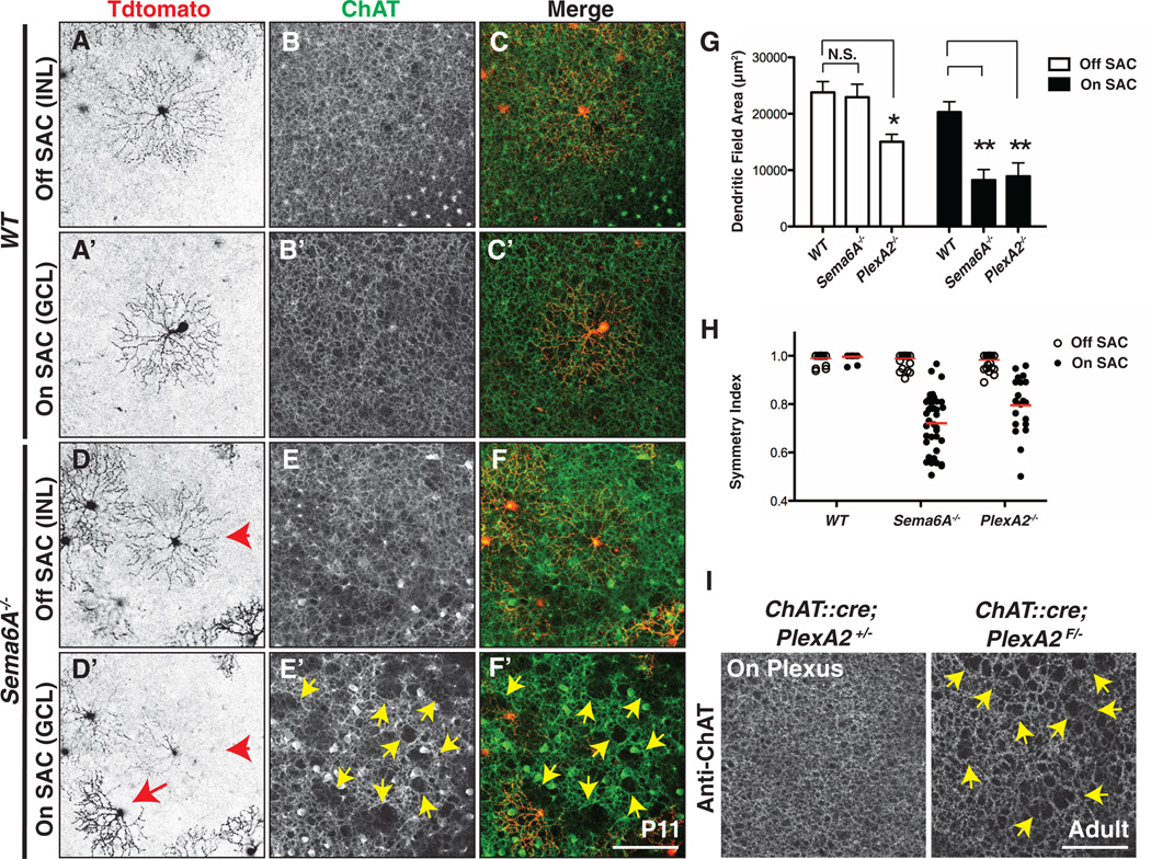
Fig. 4. Sema6A-PlexA2 signaling is dispensable for Off SAC dendritic arborization and ChAT+ plexus organization.
(A to F′) Sparse genetic labeling of On [(A′) and (D′)] and Off [(A) and (D)] SACs coupled with anti-ChAT immunostaining of On [(B′) and (E′)] and Off [(B) and (E)] SAC dendritic plexuses in the same WT [(A) to (C′)] and Sema6A−/− [(D) to (F′)] retinas. (D) to (F) and (D′) to (F′) are different focal planes from the same field. On and Off SACs in WT retinas exhibit stereotypic symmetrical morphology [(A) and (A′)]. In contrast, overall dendritic organization of Sema6A−/− On SACs [(D′), red arrow], but not Off SACs [(D) and (D′), red arrowhead], is disrupted. Similarly, SAC plexus organization revealed by anti-ChAT immunostaining shows that only the Sema6A−/− On SAC plexus is compromised, as indicated by large gaps and holes [yellow arrowheads in (E′) and (F′)]. (G and H) Quantification of dendritic field area (G) and symmetry index (H) of WT, Sema6A−/−, and PlexA2−/− On and Off SACs at P11. Red bars in (H) represent mean value for each genotype. Error bars, mean ± SD. *P < 0.01; **P < 0.001. (I) Dendritic plexus organization of On SACs in control (left) and ChAT::cre;PlexA2F/− (right) retinas. Conditional removal of PlexA2 protein in all SACs disrupts On, but not Off (see fig. S14, A and B), SAC plexus (yellow arrowheads in right panel). Scale bars, 100 µm in (F′) for (A) to (F′), 100 µm in (I) for left and right panels.

