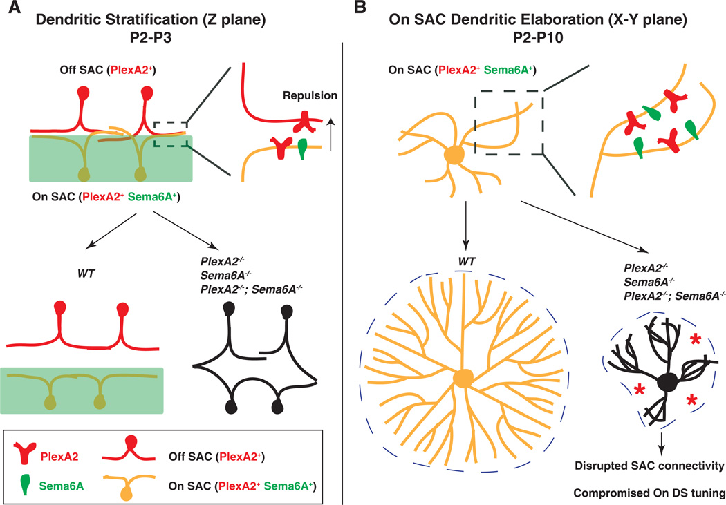
Fig. 6. Sema6A-PlexA2 signaling in the development of SACs and the functional assembly of direction-selective retinal circuits.
Sema6A-PlexA2 signaling participates in two aspects of SAC development in the early postnatal mammalian retina. (A) In the z plane, PlexA2 is expressed in both On and Off SACs, whereas Sema6A is expressed only in the lower half of the inner plexiform layer and in On, but not Off, SACs. Repulsive Sema6A-PlexA2 signaling in trans segregates initially entangled On and Off SAC processes between P2 and P3. (B) In the x-y plane, continuous expression of both Sema6A and PlexA2 in On SACs functions to separate rapidly extending dendritic processes throughout early retinal development (P2 to P10). In WT retinas, On SACs are not symmetrically organized at P0. In the presence of intact Sema6A and PlexA2 signaling, On SAC dendrites avoid each other during this early phase of SAC dendritic development, leading to elaboration of symmetric SAC dendritic fields by the end of postnatal retinal development. In the absence of Sema6A, PlexA2, or both, many On SAC dendrites fail to fully separate from one another. This affects dendritic process dynamics and eventually causes dendritic field area and symmetry defects in Sema6A−/−, PlexA2−/−, and Sema6A−/−;PlexA2−/− mutants. The disruption of On SAC dendritic plexuses affects GABA-mediated connectivity among On SACs and compromises directional tuning of On direction-selective responses.

