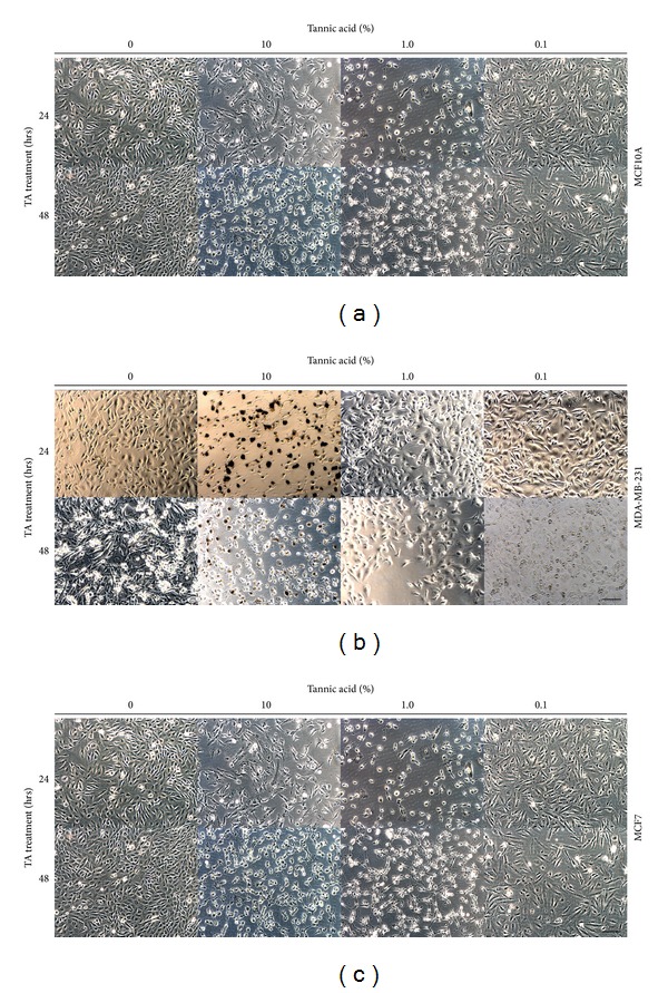
Figure 2.

Treatment of human breast cells with tannic acid. Light microscopic images of three human breast cell lines (a) MCF10A, (b) MDA-MB-231, and (c) MCF7; cells were grown to near confluence prior to treatment with 10%, 1.0%, and 0.1% TA cross-linked beads for 24–48 h. Scale bars = 50 μm.
