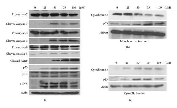
Figure 2.

Expression levels of apoptosis-related proteins were analyzed by immunoblotting. (a) Cellular proteins, (b) mitochondrial fraction, and (c) cytosolic fraction were separated by sodium dodecyl sulfate-polyacrylamide gel electrophoresis and transferred to PVDF membranes. The membranes were probed with the indicated primary antibodies and then with horseradish peroxidase conjugated goat anti-rabbit IgG. Heat shock protein (HSP)60 was used as the loading control for the mitochondrial proteins (b). Actin was used as the internal control (a) and loading control for the cytosolic proteins (c).
