
Figure 3.
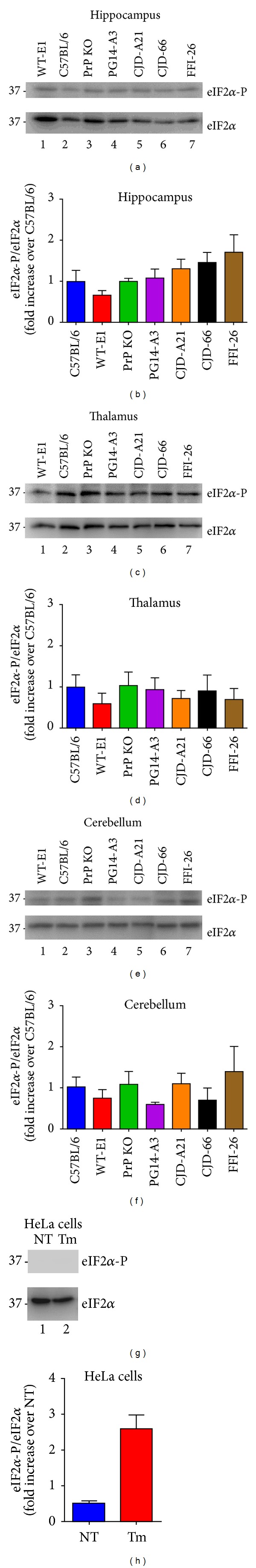
Phosphorylation of eIF2α is not increased in brains of mutant PrP mice. The same brain protein extracts (20 μg) as in Figure 2 ((a)–(f)) or lysates of HeLa cells ((g) and (h)) were analyzed by Western blot with anti-eIF2α-P and antitotal eIF2α antibodies (1 : 1000; Cell Signaling). Molecular mass markers are in kilodaltons. Phosphorylation levels were quantified by densitometric analysis of Western blots and expressed as the -fold increase over the level in C57BL/6 mice ((b), (d), (f), and (h)). Tunicamycin (Tm) treated HeLa cells were analyzed at 2 hours as control for UPR activation. Each value is the mean ± SEM of three animals of 300–350 days of age or from three independent cell preparations.