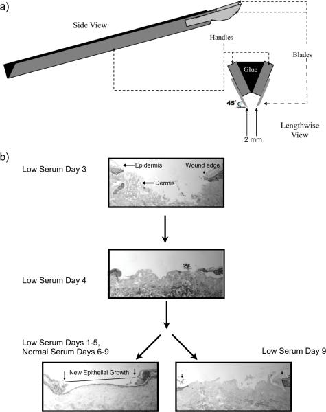
Figure 1. Linear wounds in explanted human skin.
(a) Schematic representation of the linear excision tool. Schematic representation of the linear excision tool used for creating ex-vivo linear wounds examined in this study. Two scalpel blade handles were approximated and bonded to each other with cement, blades positioned at 45° angles, and blade-to-blade width set at 2 mm. (b) Human ex-vivo linear wounds in low serum conditions maintain cell viability and re-epithelialize after the replacement of low serum media with normal serum media. Both linear and circular wounds were incubated in low serum containing media for 4 days. No evidence of reepithelialization was found in day 4 samples. Samples were then split, one group continued to receive low serum media while the other group received normal serum media up until day 9. Wounds that received normal containing media were able to fully re-epithelialize while those in low serum media remained unhealed. The wound gap was determined as the measured distance between the originally cut epithelial borders of the wound (arrows, lower left image), as visualized with H&E staining under light microscopy. The new epithelial growth is easily differentiated from the original wound margin by the absence of fully stratified epithelium and absence of the stratum corneum (outlined and easily visualized) at the wound.

