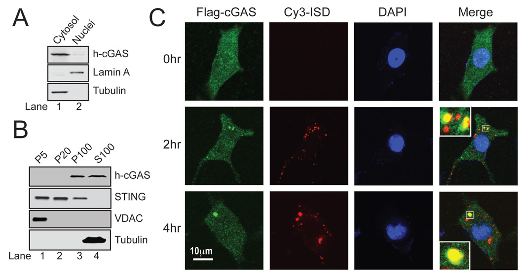
Figure 6. cGAS binds to DNA in the cytoplasm.
(A) Nuclear and cytoplasmic fractions were prepared from THP-1 cells and analyzed by immunoblotting with the indicated antibodies. (B) THP-1 cells were homogenized in hypotonic buffer and subjected to differential centrifugation. Pellets at different speeds of centrifugation (e.g, P100: pellets after 100,000 × g) and S100 were immunoblotted with the indicated antibodies. (C) L929 cells stably expressing Flag-cGAS (green) were transfected with Cy3-ISD (red). At different time points after tranfection, cells were fixed, stained with the Flag antibody or DAPI and imaged by confocal fluorescence microscopy. Inset: magnification of the area outlined in the merged images. These images are representative of at least 10 cells at each time point (representing > 50% of the cells under examination).

