
Fig. 1.
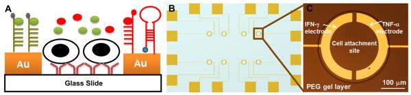
(A) Schematic illustration depicting selective modification of gold electrodes with different cytokine-binding aptamers. A pair of half ring-shaped Au electrodes fabricated on glass slides are embedded inside one PEG hydrogel and incubated with antibodies for immune cell capture. Upon injection of cells, T cells and human monocytes are bound on Abmodified glass regions. Two cytokine-binding aptamers are respectively modified on individually addressable electrodes for detecting cytokine release in real time (B) Electrode layout, where the overall device size is half of a glass slide (25 mm × 37.5 mm) (C) 300 μm diameter of PEG wells are used to capture approximately 400 cells inside one well.