
Figure 1.
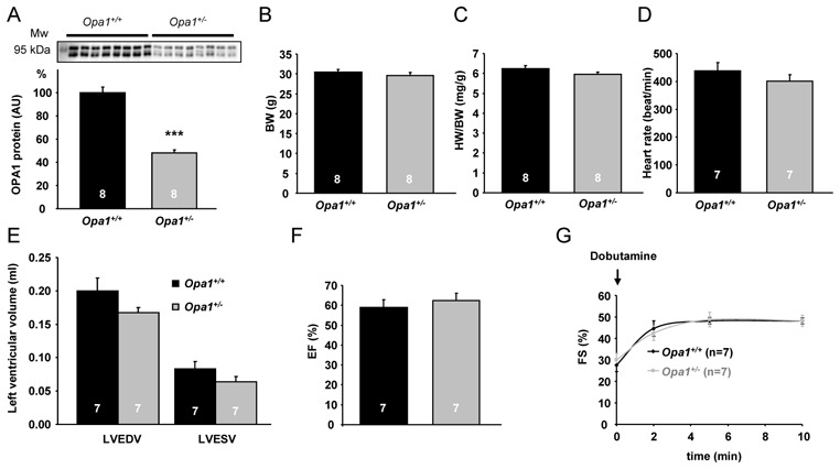
Anatomical characteristics and cardiac function of 6-month-old Opa1+/+ and Opa1+/− mice. (A) Western blot analysis of OPA1 protein. Upper panel: a representative original recording. Lower panel: mean values of OPA1 protein. ***p<0.001 versus Opa1+/+. (B) Body weight (BW). (C) Heart weight to body weight (HW/BW) ratio. (D) Heart rate. (E) LV end-diastolic volume (LVEDV) and LV end-systolic volume (LVESV). (F) LV ejection fraction (EF). (G) LV fractional shortening (FS) after dobutamine stimulation.