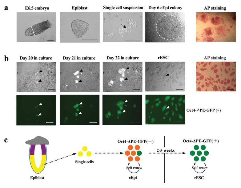
Figure 1.
Reprogramming epiblast cells from E6.5 embryos to generate rESC. (a) Derivation of cEpi from E6.5 epiblast. Epiblast tissue was divested of the proximal region (white line, panel 1) and visceral endoderm; a single cell suspension (black arrow) was cultured, which formed cEpi colonies. Note AP positive cells in cEpi in the last panel. (b) Derivation of rESC from cEpi. Note the appearance of clusters of Oct4-ΔPE-GFP-positive cells in cEpi colonies (black arrowheads), and corresponding white arrowheads for GFP in the panel below. Note that the rESC are uniformly AP positive. Scale bar: 100μm. (c) Schematic representation of reprogramming of epiblast through cEpi, and finally rESC.

