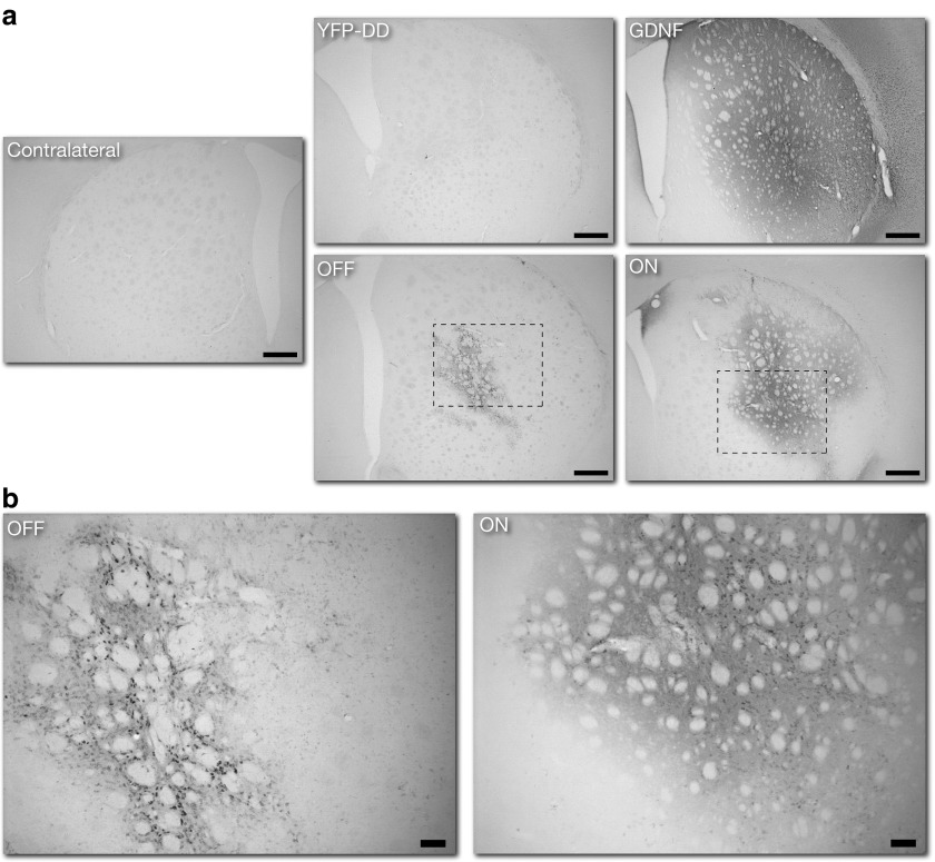
Figure 7.
Immunohistochemistry for GDNF expression in striatum. To evaluate the degree of G-F-DD expression and regulation in the 6-OHDA model, immunohistochemistry for GDNF was performed in striatal sections 6 weeks after 6-OHDA lesioning. Low magnification images of GDNF expression in striatum were performed to evaluate the general distribution of GDNF throughout the striatum (a). Scale bar: 500 µm. High magnification images were taken to highlighting the pattern of GDNF expression in the striatum of G-F-DD OFF and G-F-DD ON groups (b). Scale bar: 100 µm. DD, destabilizing domains; GDNF, glial cell line–derived neurotrophic factor.

