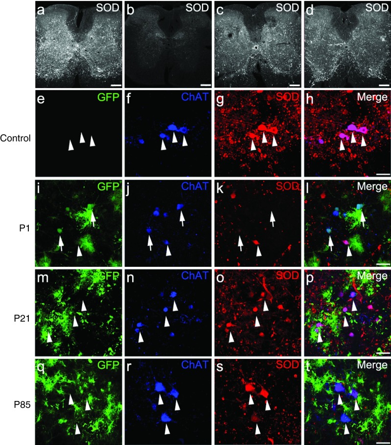
Figure 4.
Intravenous injection of AAV9-SOD1-shRNA reduces mutant protein in spinal cords of SOD1G93A mice. (a–d) Images of lumbar spinal cord sections from (a) uninjected, (b) P1-injected, (c) P21-injected, and (d) P85-injected mice were captured with identical microscope settings to qualitatively show SOD1 levels at end stage. SOD1 levels inversely correlate with survival. (e–t) Colabeling for GFP, ChAT, and SOD1 shows that AAV9-transduced motor neurons had reduced SOD1 expression (arrows) while cells that lacked GFP maintained high levels of mutant protein (arrowheads). As described in Figure 1u, higher motor neuron transduction and corresponding SOD1 reduction was observed in (i–l) P1-injected mice as compared with (m–p) P21-injected and (q–t) P85-injected mice. Bar = 100 µm. ChAT, choline acetyltransferase; GFP, green fluorescent protein; P1, postnatal day 1; P21, postnatal day 21; P85, postnatal day 85; SOD1, superoxide dismutase 1.

