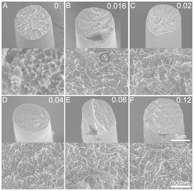
Figure 1. SEM images of the cross-sections of PEDOT:PSS/PEG-SWNTs composite fibers broken under tensile strain at low and high magnifications showing shape and microstructure of PEDOT:PSS/PEG-SWNTs composite fiber at various PEG-SWNTs loadings.
Volume fraction of PEG-SWNTs indicated at each pair images. Circle at B shows broken ends of PEG-SWNTs.

