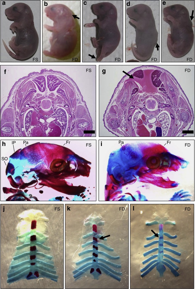
Figure 2. Paternal folate deficiency increases birth defects in offspring.
Fetuses sired by FD males displayed increased developmental abnormalities indicated by arrows (b–e) than those sired by FS males (a). Shown are the following: (b) hydrocephalus and associated craniofacial defects, (c) limb hyperextension with dysgenesis of digits, (d) spine malformation and (e) dorsal malformations. Histopathological analysis was carried out on selected FS- (f) and FD- (g) sired fetuses. (g) In this thoracic transverse section of an FD-sired fetus, the arrow indicates an imbalance between the right and left side muscular and bone tissues indicated muscular dysplasia. Scale bars, 1 mm (f,g). Skeletal staining was performed in both FS- (h,j) and FD- (i,k,l) sired fetuses. Bone is stained purple and cartilage blue. (i) FD-sired fetus lacking interparietal (IP) and supraoccipital (SO) bones and had underdeveloped frontal (Fr), parietal (Pr) bones and digits. FD-sired fetuses, with misaligned (k), or incomplete development (l) of the sternebrae plates.

