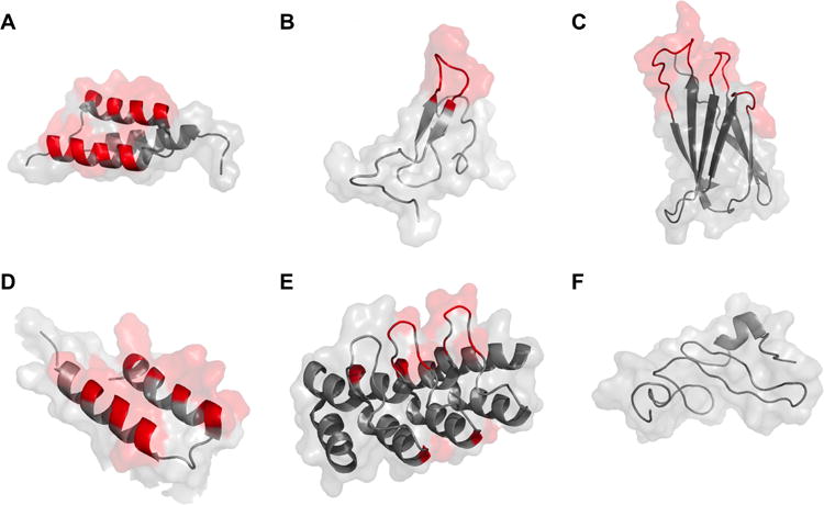
Figure 2.

Cartoon structure with surface representation of protein scaffolds used in molecular imaging applications. Variable regions are highlighted in red. Conserved portions are shown in grey. A: affibody (PDB: 2B88); B: knottin (1HYK); C: fibronectin (1TTF); D: two-helix affibody (modified from three-helix affibody 2KZJ); E: DARPin (2JAB); F: natural ligand (EGF) (2KV4).
