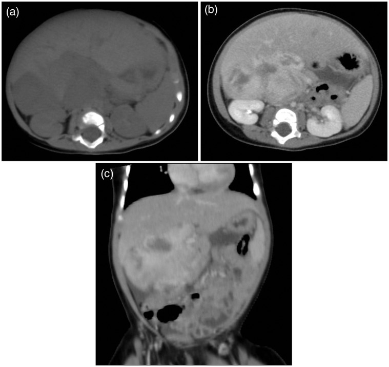
Fig. 2.
CT features of primary hepatic leiomyosarcoma. (a) Unenhanced axial CT scan of the liver revealed a large, partial demarcated and heterogeneous low attenuation mass arising from the inferior part of the right lobe of the liver without notable calcification seen. (b) Contrast-enhanced axial and (c) coronal CT scan in the venous phase showed the mass enhancing heterogeneously and intensely with areas of necrosis or hemorrhage and having mass effect to adjacent right kidney and bowel loops.

