Figure 2.
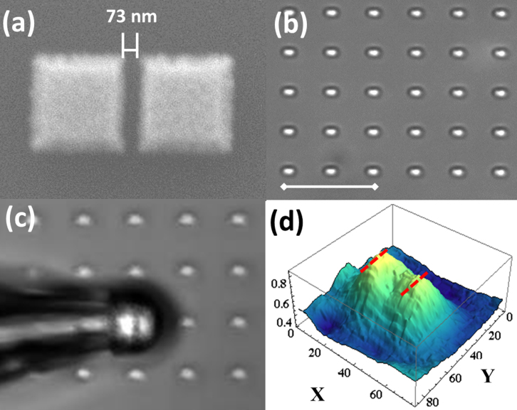
(a) SEM image of the gold split-squares nanostructure. (b) Optical microscope image of the structure in air under 50× objective lens. The scale bar is 10 μm. (c) Magnified virtual image of the structure in air by the microsphere under 50× objective lens. (d) Intensity profile of the magnified virtual image from a white dashed area in (c). X-, and Y-axes scales correspond to pixel numbers.

