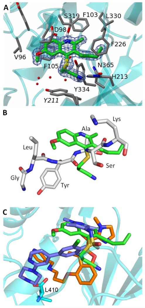
Figure 2.
A. View of 1 in chain A of PvNMT (cyan ribbon) in cylinder format and colored by atom, carbon (green) oxygen (red) nitrogen (blue) sulfur (yellow). The side chains (labeled) of surrounding protein residues are similarly colored, but with carbons in grey. Electron density (2Fo-Fc) associated with 1 contoured at 1 σ is displayed. Polar interactions with the protein and solvent are shown as dashed lines. Y211 labeled in italics is shown in two alternate conformations.
B. 1 (green) from the PvNMT ternary complex superposed with residues GLYASK of the peptide ligand from the crystal structure of the ScNMT ternary complex.9
C. 1 (green) from the PvNMT ternary complex superimposed with two inhibitors described previously for CaNMT10 (orange) and TbNMT6 (blue). The C-terminal residue of PvNMT (stick format) is labeled.

