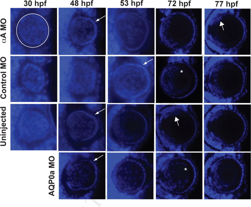
Fig. 4.

Knockdown of αA-crystallin did not inhibit fiber cell denucleation in comparison to control injected fish. Embryos were injected with either an MO that blocked the translation of αA-crystallin (αA MO), one that does not recognize any zebrafish mRNA sequence (Control MO) or one that blocks translation of a critical water channel (AQP0a MO). Other embryos were left uninjected. Embryos fixed at the specified times were cryosectioned and stained with DAPI to show cell nuclei. White circle in the upper left panel indicates the extent of the lens as a representative example with the cornea to the right. Thin arrows indicate first appearance of a noticeable lens epithelium as fiber cells become denucleated, thick arrows indicate first appearance of fiber cell nuclei restricted to a proliferative zone, and asterisks indicates residual fiber cell nuclei surrounding the lens nucleus. Times shown are hours post fertilization (hpf). At least three embryos were examined for all timepoints, except for the 77 hour timepoint, for which 2 embryos were examined.
