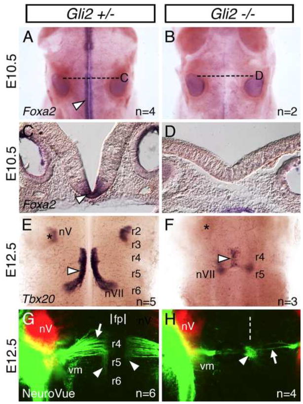
Figure 5. FBM neurons fail to migrate caudally in mouse Gli2 mutants, which lack the floor plate.
(A, B, E–H) Dorsal views of hindbrains with anterior to the top. (C, D) Cross-sections (dorsal up) at the levels indicated by broken lines (A, B). (A–D) Foxa2 in situs. In a Gli2+/− embryo (A, C), Foxa2 is expressed along the midline, corresponding to the floor plate (arrowheads), at all axial levels. In a Gli2−/− embryo (B, D), Foxa2 expression is missing, indicating the absence of a floor plate. (E–H) Tbx20 in situs (E, F) or NeuroVue injection at the cranial nerve V (red) and VII (green) exit points (G, H). In a Gli2+/− embryo (E), FBM neurons migrate caudally from r4 into r6 (arrowhead). In a Gli2−/− embryo (F), FBM neurons fail to migrate out of r4, and are found in a single cluster (arrowhead) fused across the midline. Asterisks in E, F indicate location of the trigeminal motor nucleus (nV) in r2. In a Gli2+/− embryo (G), many retrogradely-labeled FBM neurons (arrowheads) are located in r5, and the motor axons (arrow) exhibit a characteristic fan shape (genu) indicative of caudal migration. The approximate location of the floor plate (fp) is indicated. In a Gli2−/− embryo (H), FBM neurons (arrowhead) are found in a single cluster at the midline (broken line), and their axons (arrow) lack the genu reflecting the failure to migrate. The locations of the trigeminal motor (nV) and visceromotor (vm) neurons in r2 and r5, respectively, are similar between wildtype and mutant embryos.

