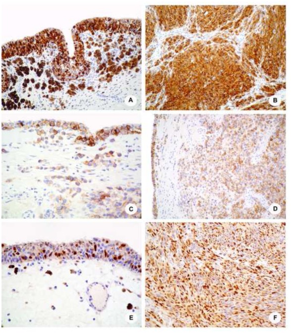
Figure 1. Immunohistochemical results.

A and B, Melan-A staining in case 17 shows a significant staining of neoplastic cells in both the in situ (A), and the invasive (B) components, with a strong and diffuse expression. The invasive components is mostly composed of sheets of spindle cells; C and D, C-Kit receptor is expressed in the majority of the neoplastic, morphologically undifferenciated cells in case 10, both by in situ (C) and invasive (D) cells; E and F, Representative cyclin-D1 immunostainings showing a strong nuclear expression by in situ and invasive components of case 17 (E) and 14 (F), respectively.
