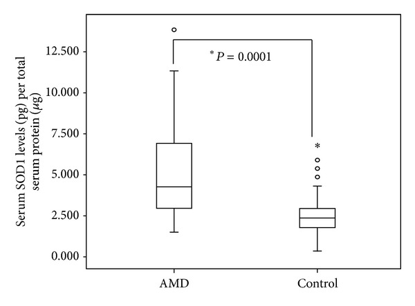
Figure 1.

Serum levels of SOD1 in AMD and normal controls. Boxes include values from the first quartile (25th percentile) to third quartile (75th percentile). The thick horizontal line in the box represents median for each dataset. Outliers and extreme values are shown in circles and asterisk, respectively. Levels of SOD1 were normalized to total protein. Data was analyzed by using the Mann-Whitney U test.
