
Figure 2. Laforin antibody selection.
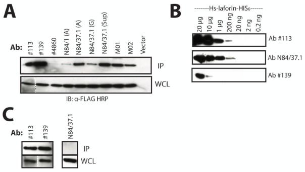
A. Immunoprecipitation of overexpressed laforin using available antibodies. FLAG-laforin was immunoprecipitated using a variety of α-laforin antibodies in conjunction with Protein A Sepharose. We tested protein A-purified rabbit polyclonal α-laforin antibodies #113, #139, #4860, protein A- and G-purified mouse monoclonal α-laforin antibodies N84/1 and N84/37.1 from NeuroMabs, and several commercially available mouse monoclonal α-laforin antibodies (M01 and M02, Abnova). “(A)” or “(G)” indicates affinity purification of an antibody with either protein A or protein G, respectively. “Sup” indicates unpurified tissue culture supernatant. Cells containing empty vector were immunoprecipitated with α-FLAG agarose. The depicted image is a representation. The dotted line indicates where an image portion is a composite of the same image due to the presence of molecular weight marker in between the lanes. B. Detection limit of laforin antibodies. Serial dilutions of recombinant laforin (20.0 μg to 0.2 ng) were probed with the α-laforin antibodies. Only α-laforin antibodies #113, N84/37.1, and #139 are shown, as they displayed the most sensitive detection of recombinant laforin. Primary antibody was used at 1:1000 and secondary antibody at 1:3000. The depicted image is a representation. C. Immunoprecipitation of endogenous laforin from HepG2 cultures. Only the polyclonal α-laforin antibodies #113 and #139 were able to immunoprecipitate endogenous laforin. While the other antibodies did not immunoprecipitate laforin, laforin was detected in their WCL samples. Only the result from α-laforin antibody N84/37.1 is shown to depict this negative result. The depicted image is a representation. The dotted line indicates where an image portion is a composite of the same image due to the presence of molecular weight marker in between lanes. The above experiments were performed a minimum of three times.