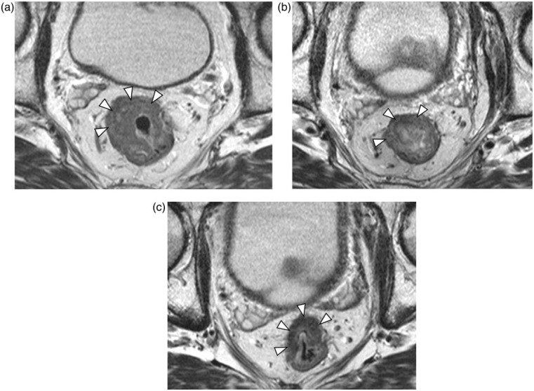
Figure 1.
Images of 59-year-old man with rectal cancer. Axial fast spin-echo T2-weighted magnetic resonance (MR) imaging (a) pretherapy, (b) at the end of the second week of chemoradiotherapy (CRT), and (c) after CRT and before surgery, showing a volume reduction of 45%. Arrowheads indicate the tumor outline.

