
Figure 10.
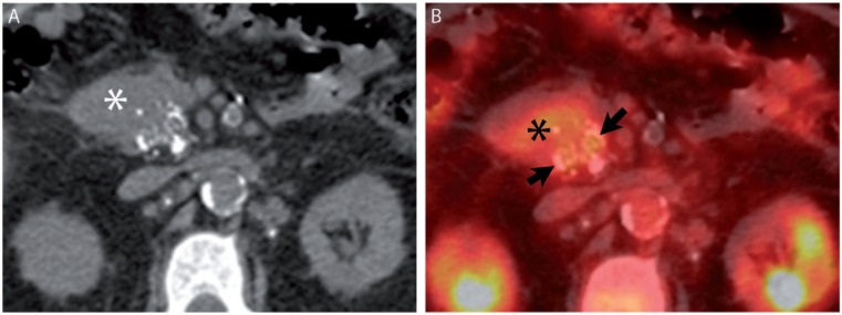
A 68-year-old man with pancreatic ductal adenocarcinoma. (A) Axial noncontrast CT image from [18F]fluorodeoxyglucose (FDG)-positron emission tomography (PET)/CT demonstrates stippled calcification in a pancreatic head mass (asterisk). (B) Fused image from FDG-PET/CT demonstrates FDG avidity engulfing the calcifications (arrows).