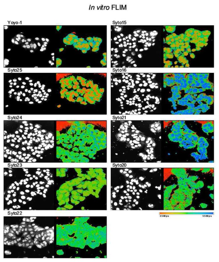
Figure 4.

In vitro Fluorescent Lifetime Imaging Microscopy (FLIM): Discrimination of cellular DNA and RNA. Intensity (left panel) and lifetime images (right panel) of MCF-7 cells stained with a range of green-fluorescent nucleic acid binding dyes. The dyes are commonly used as nuclear or cell counter stains in standard fluorescent microscopy (left panel) but are known to differ from each other in one or more characteristics, including DNA/RNA binding selectivity. Here, additional contrast provided by FLIM of Syto15 and Yoyo-1 distinguished cytoplasm, nucleus and nucleoli. Contrast observed with Yoyo-1 is due to longer lifetime of dye bound to RNA and shorter lifetime of dye bound to DNA (data not shown). FLIM contrast observed with other dyes was either minimal (Syto23) or offered less contrast across cytoplasmic, nuclear and nucleolar compartments (Syto25, Syto16, Syto21, Syto22, Syto20). This study identified dyes appropriate for rapid cell image segmentation using FLIM rather than requiring dye multiplexing in either live or fixed cells and suggests that additional screening to identify FLIM contrast for biological event monitoring e.g. apoptosis or cellular differentiation may be worthwhile. Images acquired by Clifford Talbot et al. in the Photonics Group at Imperial College London using a laser scanning confocal TCSPC microscope.
