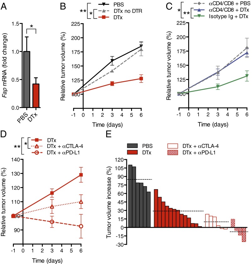
Fig. 3.
Conditional depletion of FAP+ stromal cells and immune control of PDA. (A) KPCD mice with PDA received DTx or PBS; after 6 d, tumoral Fap mRNA was measured by quantitative RT-PCR (qRT-PCR) (PBS, n = 5; DTx, n = 7). (B) KPC mice with or without the DTR BAC transgene received DTx or PBS, and tumor volumes were measured by ultrasound (PBS, n = 6; DTx, n = 8; DTx to non-DTR transgenic, n = 4). (C) KPCD mice with PDA received CD4- and CD8-depleting antibodies or control IgG before and during treatment with DTx or PBS. Tumor volumes were measured (α-CD4/8 + PBS, n = 3; α-CD4/8 + DTx, n = 5; isotype IgG + DTx, n = 5). (D) KPCD mice with PDA received α-CTLA-4 or α-PD-L1 during treatment with DTx or PBS, and tumor volumes were measured [DTx, n = 13 (representing all DTx-treated KPCD mice presented in B and C); α-CTLA-4 + DTx, n = 6; α-PD-L1 + DTx, n = 4]. (E) Waterfall plots demonstrate the final tumor volume changes in individual mice. Dashed lines indicate the mean tumor volume for each treatment group. *P < 0.05; **P < 0.01.

