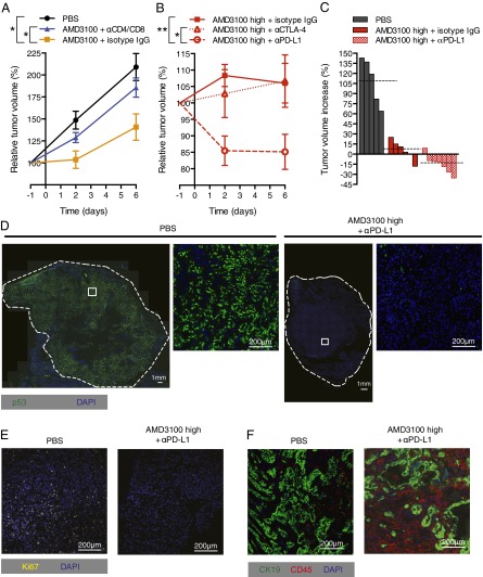
Fig. 5.
Inhibition of CXCR4 by AMD3100 and immune elimination of PDA cells. (A) PDA-bearing mice, some of which had been pretreated with depleting CD4 and CD8 antibodies or control IgG, received PBS or AMD3100 by continuous infusion, and tumor volumes were measured by ultrasound (PBS, n = 5; AMD3100 + isotype IgG, n = 6; AMD3100 + α-CD4/8, n = 4). (B) PBS or AMD3100 (high dose) was given to PDA-bearing mice that were treated with α-CTLA-4, α-PD-L1, or control IgG (AMD3100 high + α-CTLA-4, n = 4; AMD3100 high + α-PD-L1, n = 7; AMD3100 high + isotype IgG, n = 6). (C) Waterfall plots present the final changes in tumor volumes in individual mice from A and B. Dashed lines indicate the mean tumor volume for each treatment group. (D) Tissue sections from PDA tumors taken from mice treated with PBS or AMD3100 high + α-PD-L1 for 6 d were stained for p53, and the entire cross-sections were imaged. White squares show the regions corresponding to the magnified images. Tissue sections from PDA tumors of mice treated with AMD3100 high + α-PD-L1 or PBS for 6 d were stained for Ki67 (E) or CD45 and CK19 (F) and analyzed by IF microscopy. *P < 0.05; **P < 0.01.

