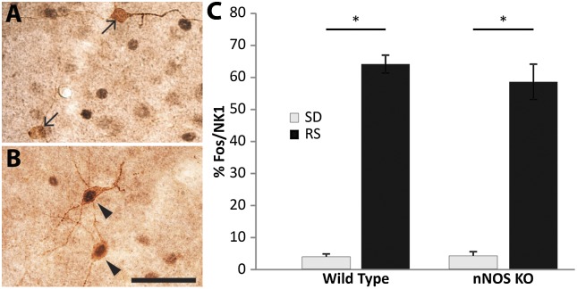
Fig. 6.
Cortical NK1 neurons are activated during RS in the absence of nNOS. (A) Cortical NK1 neurons (brown) do not express Fos (black) in nNOS KO mice killed at the end of 6 h SD. (B) In contrast, cortical NK1 neurons express Fos after 6 h SD followed by a 2 h RS opportunity. (C) The %Fos/NK1 neurons is significantly increased after 2 h RS in both WT and KO mice, even though the homeostatic response differs between these two strains as demonstrated in Fig. 5. Arrows, single-labeled NK1 neurons; arrowheads, double-labeled Fos/NK1 cells. (Scale bar, 50 µM.)

