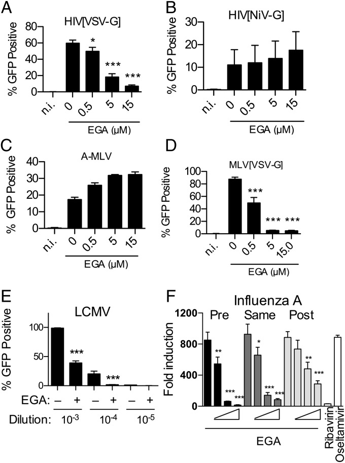
Fig. 6.
EGA inhibits entry of low pH-dependent viruses. (A and B) EGA inhibits transduction by lentiviral particles pseudotyped with (A) VSV-G but not (B) NiV envelope proteins. (C and D) EGA does not inhibit infection by (C) A-MLV, but does inhibit transduction by (D) MLV particles pseudotyped with VSV-G. (A–D) HeLa cells were incubated with indicated dose of EGA for 1 h and then challenged with GFP-encoding viral particles for 2 d, followed by quantification of GFP expression by flow cytometry. Cells that were not infected (n.i.) did not express GFP. (E) Vero cells were incubated with 20 µM EGA for 1 h and then challenged with a GFP-encoding variant of the Armstrong strain of LCMV at three dilutions as shown. Two days later, cells were fixed and analyzed by flow cytometery. (A–E) Average percentage of GFP-expressing cells from three independent experiments shown ±SD. (F) HeLa cells transfected with a Flu gLuc reporter were incubated with EGA (0.5, 5, or 15 µM) 1 h pre, simultaneously, or 1 h post-challenge with influenza A/WSN/33. The following day, luciferase activity was measured and normalized to uninfected controls. Average fold luciferase induction from three independent experiments is shown ±SD.

