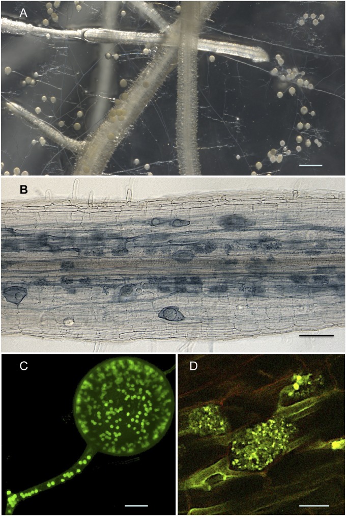
Fig. 1.
(A) In vitro coculture of R. irregularis with carrot roots showing extraradical hyphae and spores. (Scale bar: 750 µm.) (B) Colonized carrot root showing fungal colonization that is restricted to the root cortex where the fungus produces vesicles and/or intraradical spores, and arbuscules. (Scale bar: 100 µm.) (C) Typical multinucleated asexual spore of R. irregularis and its attached coenocytic hyphae observed by confocal laser scanning microscopy. Nuclei were stained using SYTO Green fluorescent dye and are shown as green spots. (Scale bar: 10 µm.) (D) Arbuscules, highly branched structures formed by the fungus inside cortical cells, are considered the main site for nutrient exchanges between the mycorrhizal partners. The yellow-green fungal structures are detected by wheat germ agglutinin-FITC labeling on root sections, whereas PCWs are shown in red. (Scale bar: 10 µm.)

