Figure 1.
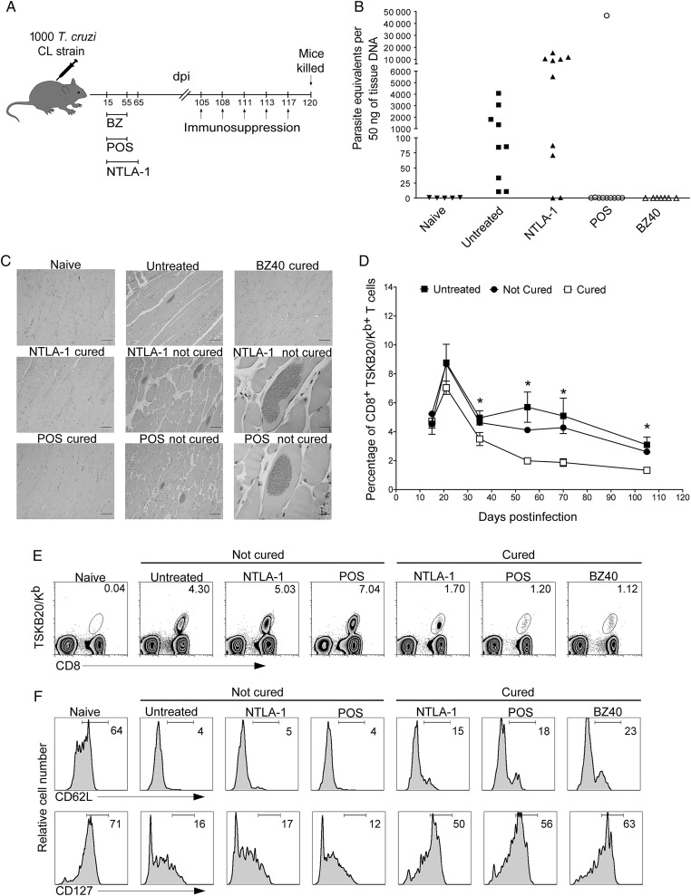
A shift to a TCM phenotype in the Trypanosoma cruzi–specific CD8+ T cells is diagnostic of cure in T. cruzi infection. A, Schematic representation of infection, treatment, and immunosuppression. B, T. cruzi DNA isolated from skeletal muscle tissues of untreated or treated mice and suppressed with cyclophosphamide (15 days after suppression started) determined by quantitative real time polymerase chain reaction. C, Histological sections of the skeletal muscle at 120 days postinfection of naive, untreated, benznidazole 40-day–, posaconazole–, and NTLA-1–treated mice that were cyclophosphamide suppressed. Scale bar represents 200 µm in the left and middle columns and in the upper panel of the right column. In the right column middle and bottom row scale bar represents 20 µm. D and E, The MHC-peptide tetramer of the immunodominant TSKB20/Kb epitope was used to detect T. cruzi–specific CD8+ T cells in the blood of untreated (filled squares) and treated mice (cured: open square; not cured: filled circles) during the evolution of the infection (D) and at 105 days postinfection before the immunosuppression (E). Data in (D) are shown as mean ± standard error of the mean. *P < .05 between cured and not cured groups or cured and untreated groups. Numbers in (E) indicate the percentage of tetramer+ cells among the CD8+ T-cells population. F, Expression of classical memory (CD62L) and memory maintenance (CD127) markers in blood on the CD8+ T cells (naive) and on the CD8+ TSKB20-tetramer+ T cells from untreated and treated mice at 105 days postinfection. Data are representative of 2 independent experiments with 5–10 mice per group. Abbreviations: BZ, benznidazole; dpi, days postinfection; POS, posaconazole; T. cruzi, Trypanosoma cruzi.

