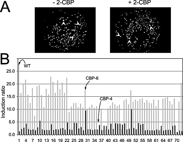
Figure 5.

Purification of HbpR mutants responsive to 2‐CBP. A. Fluorescence from E. coliFACS‐enriched mutant pool colonies (left, constitutive mutants) or after exposition to 2‐CBP on filter membrane (right). Arrows point to particularly bright fluorescing colonies such as selected for further study. B. Fluorimetry analysis of some 70 mutants purified via FACS and fluorescence plates after exposure to 2‐HBP (light grey bars) or 2‐CBP (dark grey) at 20 and 100 µM respectively. The ordinate shows induction factor compared with cells in buffer only. Culture number 1 and 73 are expressing HbpR wild type.
