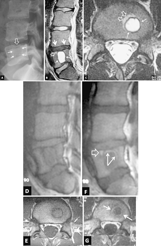
Figure 1.

Imaging 1 week after injury. (A) The lateral plain film shows a lytic lesion with a thin sclerotic border (arrows). A focal depression is also evident in the L5 upper epiphyseal plate (open arrow). The sagittal (B) and axial (C) T2-weighted magnetic resonance (MR) images show a large cystic area with a low signal intensity border corresponding to a giant Schmorl's node (arrows). Reactive bone marrow edema is seen anterior to the lesion (open arrows). The L4–L5 intervertebral disc is degenerated (thick arrowheads). The sagittal (D) and axial (E) T1-weighted MR images and the corresponding images following intravenous contrast medium administration (F, G) show a “ringlike” enhancement within the sclerotic border (arrows) and anterior to the lesion (open arrow).
