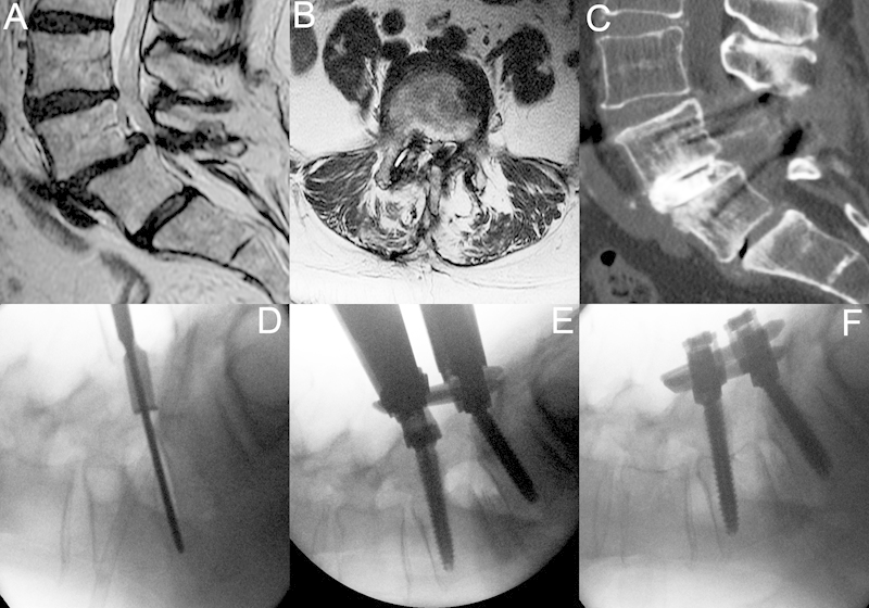
Figure 4.

A 55 year old man with history of back pain and neurogenic claudication. This patient had failed a previous laminectomy and further nonoperative treatment and underwent a minimally invasive lumbar redo laminectomy, discectomy, interbody fusion, and instrumentation through a 22-mm tubular retractor. (A, B) Lumbar magnetic resonance imaging shows grade I/II spondylolisthesis with severe stenosis. (C) Postoperative computed tomography 18 months after surgery. (D) Lateral X-ray on the operating room table reveals a grade II spondylolisthesis. A 22-mm tubular retractor is in place, and the disc space is entered and discectomy is performed. (E, F) An expandable cage has been inserted and bone graft has been placed. Instrumentation has been placed and the spondylolisthesis is reduced by locking down the L5 cap and reducing L4.
