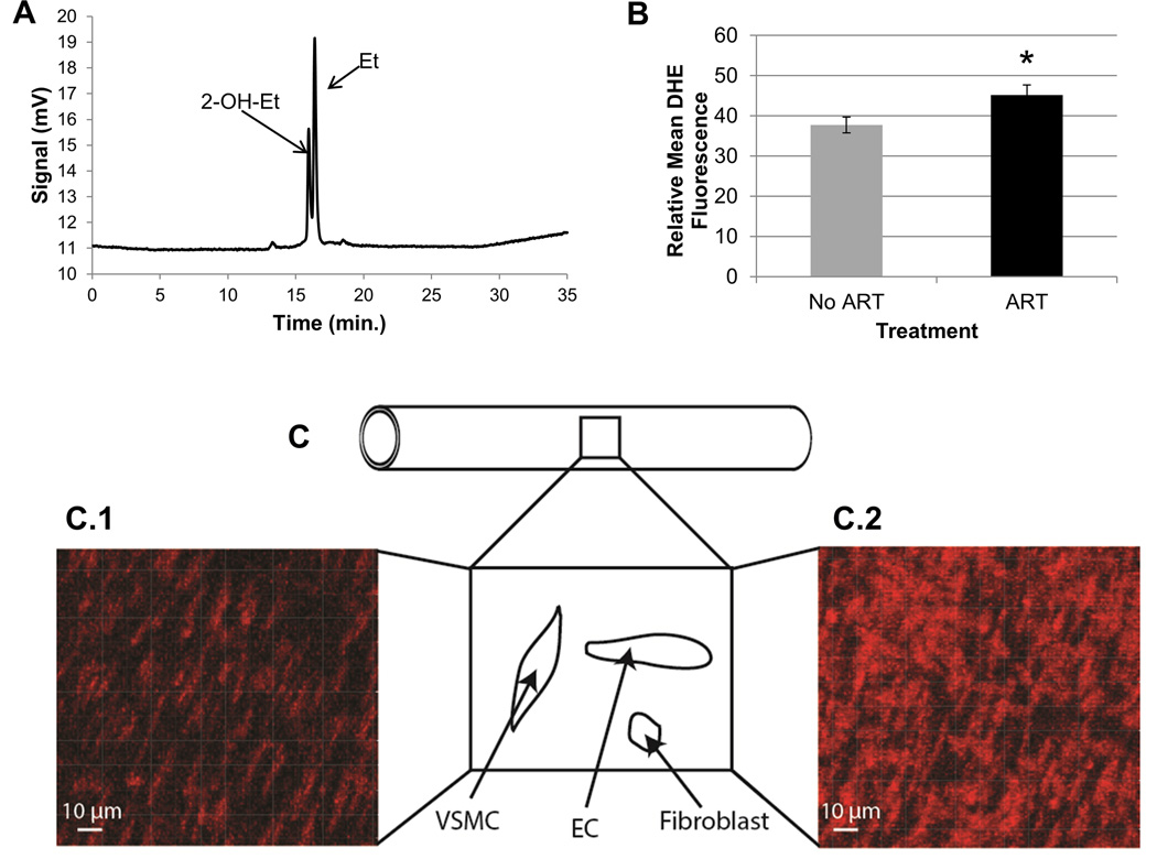
Figure 4. Detection of extracellular and intracellular ROS in mesenteric resistance arteries of offspring.
A. Two fluorescent byproducts of dihydroethidium (DHE) were detected by high performance liquid chromatography (HPLC) in offspring (55 ± 0.5 days) mesenteric resistance arteries. Resistance arteries were incubated with 50 µM DHE for one hour. 2-hydroxyethidium (2-OH-Et; specific byproduct of superoxide and DHE) eluted at 16.19 ± 0.03 min and the ethidium (Et; less specific byproduct) eluted at 16.61 ± 0.03 min. Areas under each curve were determined using the Empower program (Waters, Build #1154) and were normalized to the concentration of protein in each vessel before comparisons between treatments. Protein amounts were with a Micro BCA™ Protein Assay Kit. B. Determination of levels of intracellular ROS in offspring mesenteric resistance arteries using confocal microscopy for relative DHE fluorescence. Intracellular DHE fluorescence (red) was measured by confocal microscopy. Gain was set at 800 V. Data represent the group average ± S.E.M. of DHE fluorescence over the average of two areas of the mesenteric resistance arteries for offspring conceived naturally (No ART; gray, n=32) or with the use of ART (ART; black, n=31). C. Shown is a diagrammatic representation of a resistance artery indicating that a portion of the middle section of the vessel was flattened and the vascular wall imaged using confocal microscopy to detect DHE-dependent fluorescence. Within the wall nuclei of different cells (i.e., VSMC=vascular smooth muscle cells, EC=endothelial cells, and fibroblasts) bind to the dye and highlight its fluorescence intensity. Notice that the nuclei for different cells have specific morphologies and spatial orientations. The micrograph of a longitudinal section of a mesenteric resistance artery from a naturally conceived LF male is shown in C.1, and an ART conceived LF male in C.2. Red fluorescence results from the oxidation of DHE. Fluorescence was quantified with the Imaris 7.6.1 software. ART = assisted reproductive technologies. DHE = dihydroethidium. ROS = reactive oxygen species. *p=0.056.

