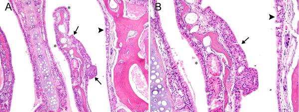
Figure 6. Respiratory/transitional epithelial hyperplasia.

A) Epithelial hyperplasia, with transitional features (arrows) forms a plaque-like thickening of the epithelial lining on the lateral side of a nasoturbinate in Level II of the same female mouse with the adenoma in Level I (2-year studies) that is shown in Fig. 4. Respiratory hyperplasia is also noted on the lateral wall (arrowhead). Normal appearing respiratory epithelium is indicated by asterisks for comparison. B) The stratified, non-ciliated epithelium with transitional features (arrow), and the hyperplastic, ciliated epithelium with respiratory epithelial features (arrowhead) are better shown in this higher magnification image. The more normal columnar, ciliated respiratory epithelium is marked with an asterisk for comparison. Original objective magnification: A) 10×, B) 20×.
