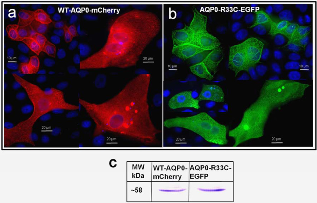
Fig. 3.
Localization and colocalization of WT-AQP0-mCherry and AQP0-R33C-EGFP. (a, b) Epifluorescent images of MDCK cells transfected with the WT-AQP0-mCherry and AQP0-R33C-EGFP, respectively. c. Western blot analysis of MDCK cells expressing WT-AQP0-mCherry (lane 1) and AQP0-R33C-EGFP (lane 2). (d–f) Cells cotransfected with WT-AQP0-mCherry and AQP0-R33C-EGFP constructs; (d) cotransfected cell viewed under mCherry fluorescent filter; (e) the same cells under an EGFP fluorescent filter; (f) overlaid image of (d) and (e).


