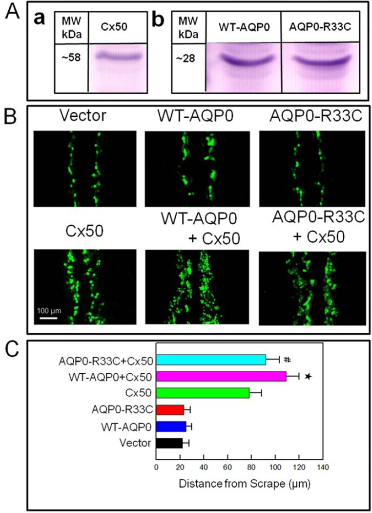
Fig. 7.
Scrape-loading dye transfer assay showing AQP0-facilitated gap junction coupling. (A). Western blotting of (a) Cx50, (b) WT-AQP0 and AQP0-R33C proteins to show expression of the respective proteins on L-cells. (B). Lucifer yellow (LY) dye transfer in L-cells stably expressing vector, WT-AQP0, AQP0-R33C, Cx50, WT-AQP0 + Cx50 or AQP0-R33C + Cx50. The extent of dye transfer was quantified by measuring distance from the scrape line to the dye front of LY. (C). Bar graph representing the data collected, presented as mean ± SD. (n=5). * Increase in gap junction coupling in WT-AQP0 + Cx50 transfected cells was statistically significant (P < 0.001) compared to Cx50 transfected cells; # Reduction in gap junction coupling in AQP0-R33C + Cx50 transfected cells was statistically significant (P < 0.001) compared to WT-AQP0 + Cx50 transfected cells.

