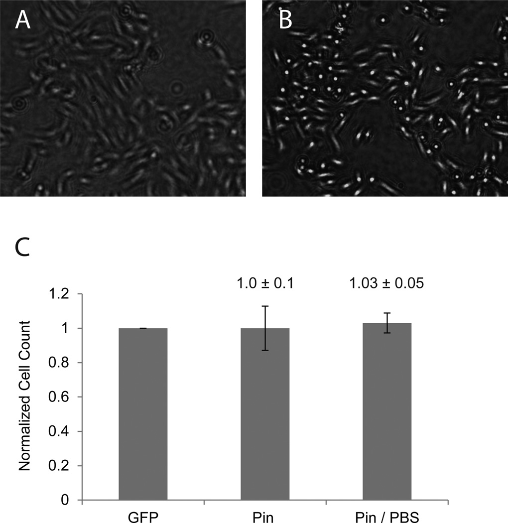
Figure 2. Cell counting after swelling by PBS treatment.
SHEP-GFP cells were seeded at 1500k in a 12 well plate and grown for 2 days with a media switch after 24 hours. Pinhole illuminated bright-field images were recorded in cell media (A). In order to increase contrast in images complete media was exchanged for PBS. Cells were immersed in PBS for 15 minutes before imaging (B). Cells were counted as described in Fig. 1. Cell counts of pinhole illuminated images with and without PBS incubation were normalized against fluorescence images of the same viewing field (means ± SD, n = 9).

