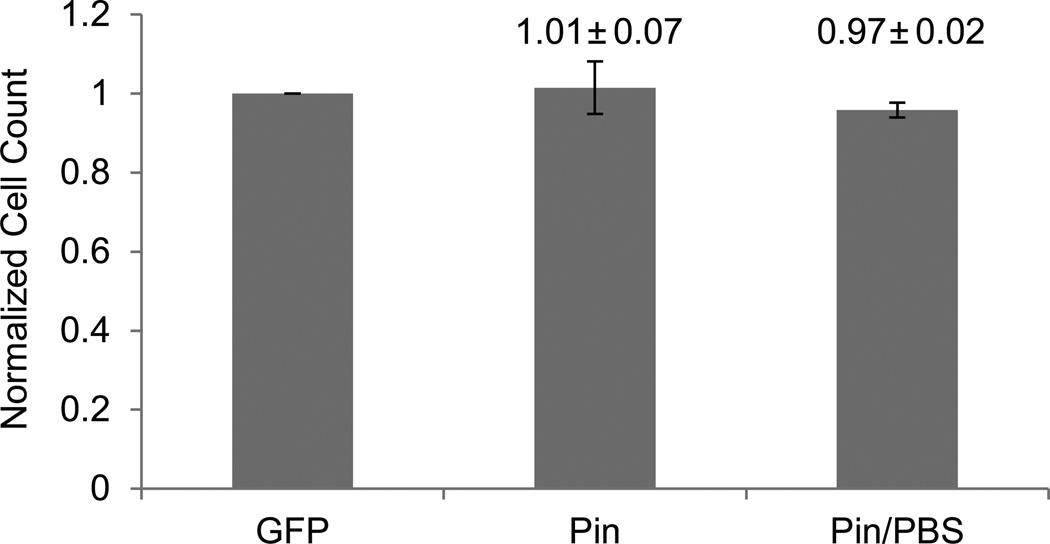
Figure 4. Counting of dense confluent cell layers.
SHEP-GFP cells were seeded at very high cell densities of ~5000k / well and allowed to grow for one day, resulting in a fully confluent cell layer. Cells were imaged with fluorescence, pinhole, and pinhole + PBS techniques. Cell counts of pinhole-illuminated images with and without PBS incubation were normalized against fluorescence images of the same viewing field (means ± SD, n = 6).

