
Fig. 3.
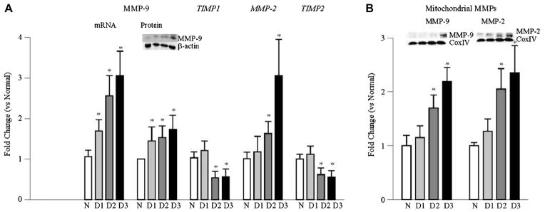
Effect of duration of diabetes in rats on retinal MMP-9 and MMP2: (A) Gene expressions of MMP-9, MMP-2 and their intracellular inhibitors TIMP1 and TIMP2 respectively, were quantified by real time qPCR in the retina from rats diabetic from 15 days to 6–12 months using gene specific rat primers and β-actin as an internal control. Protein expression of MMP-9 was quantified by western blot technique. (B) Accumulation of MMP-9 and MMP-2 in the mitochondria was quantified by western blot technique using Cox IV as loading control. Each measurement was made in 6–12 rats/group, and the values are represented as mean ± SD *p < 0.05 compared to normal. Values obtained from normal rats are considered as 1. N = normal, D1, D2 and D3 = rats diabetic for 15 days, 2 months and 6–12 months respectively.