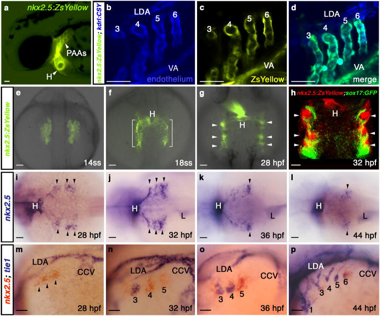
Figure 1. nkx2.5 is expressed in presumptive PAA endothelial progenitors.
a, Tg(nkx2.5:ZsYellow) embryo at 60 hpf exhibiting fluorescence in the heart and PAAs. b-d, Tg(nkx2.5:ZsYellow); Tg(kdrl:CSY) embryo at 60 hpf with overlapping yellow and blue fluorescence in the endothelium of PAAs 3-6 and the VA. The LDA exhibits blue fluorescence exclusively. e-g, ZsYellow fluorescence in Tg(nkx2.5:ZsYellow) embryos at 14ss (e), 18ss (f), and 28 hpf (g). Brackets and arrowheads highlight non-cardiogenic nkx2.5+ cells in the pharynx. h, Tg(nkx2.5:ZsYellow); Tg(sox17:GFP) embryo at 32 hpf with nkx2.5+ pharyngeal cells (red) and sox17+ pharyngeal endoderm (green); i-l, in situ hybridization time-course of nkx2.5 expression in the pharynx. Arrowheads mark nkx2.5+ pharyngeal clusters. m-p, Double in situ hybridization time-course of nkx2.5 (red) and tie1 (blue) expression. PAA1 (labelled in p) forms earlier in development and never expresses nkx2.5. tie1+ clusters are numbered according to the mature PAA they derive. a-d, m-p, lateral views, anterior left; e-h, dorsal views, anterior up; i-l, dorsal views, anterior left; a-p, n>20 embryos per group. Abbr: VA, ventral aorta; LDA, lateral dorsal aorta; ss, somite-stage; hpf, hours post-fertilization; H, heart; L, liver; CCV, common cardinal vein.

