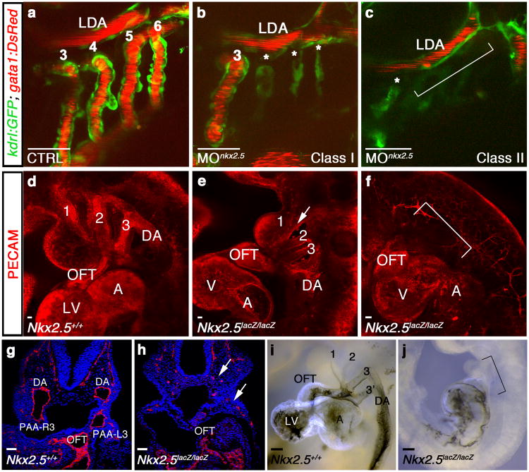
Figure 3. Nkx2.5 is required for vertebrate PAA formation.
a-c, Tg(kdrl:GFP); Tg(gata1:DsRed) zebrafish embryos with green endothelial cells and red erythrocytes at 60 hpf. a, Control (CTRL) embryo with patent PAAs 3-6 and LDA (n=60/60). b, Class I nkx2.5 morphant (MOnkx2.5) in which one (shown) or two PAAs support blood flow. Asterisks label malformed PAAs (n=88/116). c, Class II nkx2.5 morphant lacking patent PAAs (bracket, n=22/116). d-f, Whole mount control (d) or Nkx2-5 null (e,f) mouse embryos stained for PECAM1 (red) at E9.5 (n=5 per group). Nkx2-5 null embryos displayed disrupted PAAs 1-3 with residual isolated endothelial cells (arrow, e) or a complete absence of PAAs altogether (bracket, f). g,h, Sections through control and Nkx2-5 null embryos stained with PECAM (red) and DAPI (blue). Arrow shows residual endothelial cells (h). i,j, Ink injections in control and Nkx2-5 null animals at E9.5 (n=5 per group). Bracket in j indicates absence of flow through the mutant OFTs. a-c, lateral views, anterior left; d-f, i,j, lateral views, anterior up; g,h, coronal sections. Scale bar = 50μm. Abbr: LDA, lateral dorsal aorta; DA, dorsal aorta; OFT, outflow tract; LV, left ventricle; A, atrium; V, ventricle; PAA number and left (L) and right (R) designations indicated.

