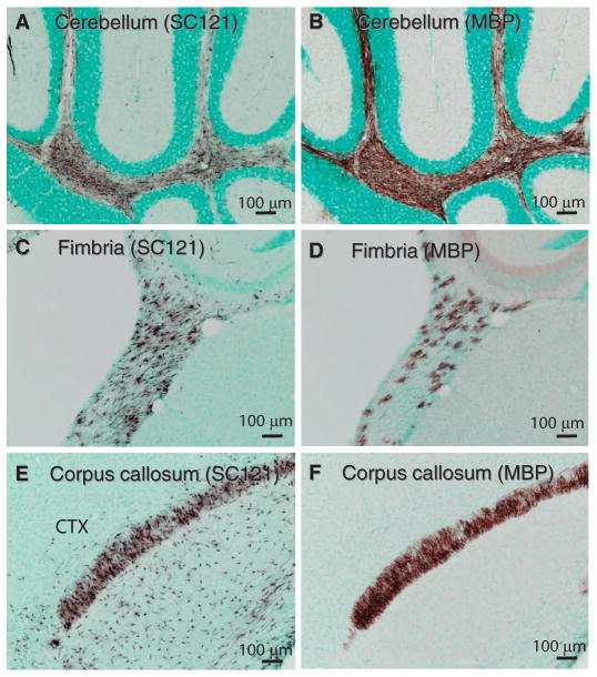
Fig. 4.
Production of myelin after HuCNS-SC transplant into Shi-id mice. Representative images from neonatal Shi-id mice after HuCNS-SC transplantation. (A) SC121 staining (brown) revealed diffuse human stem cell engraftment in cerebellar white matter 9 weeks after transplant. (B) MBP staining (brown) in an adjacent section of the cerebellum revealed extensive myelination. (C and E) SC121 staining (brown) revealed diffuse engraftment of HuCNS-SCs in the fimbria of the fornix (C) and in the cortex (CTX) and corpus callosum (E) at 12 weeks after transplant. (D and F) Corresponding MBP staining in the fimbria of the fornix and in the cortex and corpus callosum. Regional boundaries are delineated with methyl green counterstain.

