
Fig. 5.
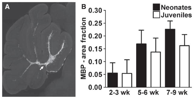
Oligodendrocyte differentiation and myelination after HuCNS-SC transplant. (A) A representative section showing MBP staining of cerebellar white matter tracts 6 weeks after transplant in a neonatal animal (arrow). (B) Quantitation of MBP staining of cerebellar white matter in transplanted neonatal and juvenile mice over time. There were no significant differences in the magnitude of MBP staining between neonates and juveniles at any time point. Neonate: n = 2 (2 to 3 weeks), n = 6 (5 to 6 weeks), n = 3 (7 to 9 weeks); juvenile: n = 3 (2 to 3 weeks), n = 5 (5 to 6 weeks), n = 5 (7 to 9 weeks).