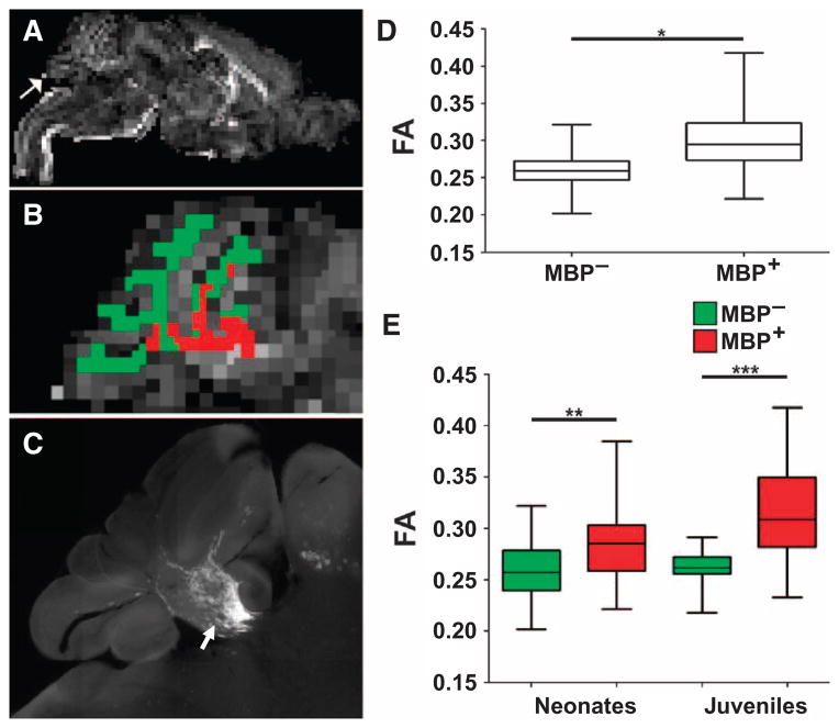
Fig. 8.
High-field MRI analysis of HuCNS-SC–derived myelination. Human myelination after HuCNS-SC transplant into Shi-id mice resulted in microstructural changes in cerebellar white matter tracts identified as an increase in FA. (A) Representative low-power parasagittal FA map at one level of the entire brain from a neonatal animal at 6 weeks after transplant. Arrow indicates the cerebellar region shown in (B). (B) Detailed view of the FA map of the cerebellum. A white matter segmentation for the FA map was generated based on the corresponding T2-weighted image and was used to identify the regions of cerebellar white matter that corresponded to human myelination (red) or to an absence of myelin (green). (C) A high-resolution image of cerebellar MBP staining (arrow) at the level of the FA maps in (A) and (B). (D) FA data from regions of white matter segmentation corresponding to myelin (MBP+) or absence of myelin (MBP−). Data are derived from MRI analyses of neonatal (n = 4) and juvenile (n = 4) transplanted Shi-id mice. (E) There was an increase in FA in regions of human myelination (MBP+; red) relative to regions without myelin (MBP−; green) in both neonatal and juvenile transplanted mice (*P < 0.0001; F = 56; mixed-effects model; **P < 0.047, ***P < 0.001).

