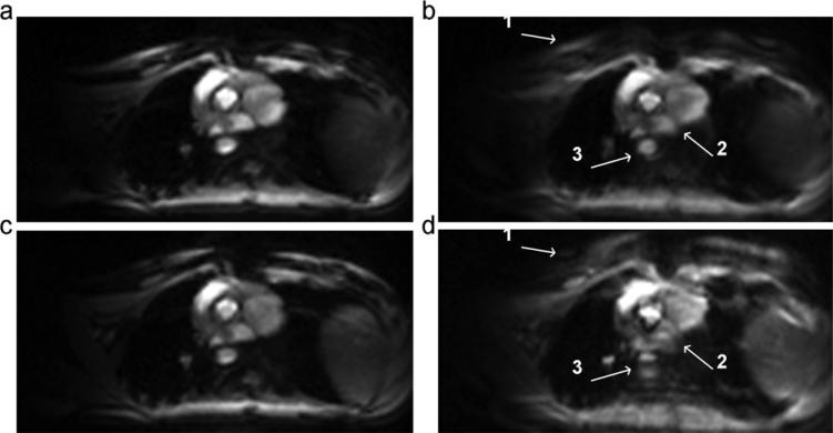
Figure 6.
Images for top-down (a, b) and center-out trajectory (c, d) for an off-resonance frequency of 0 Hz (a, c) and 100 Hz (b, d). White arrows in image acquired with center-out trajectory (d) point to artifacts caused by splitting and blurring at the (1) chest wall, (2) boundary between the right and left atrium, and (3) the descending aorta not seen in images acquired with top-down trajectory (b). While this 100 Hz off-resonance frequency was artificially introduced for demonstration purposes, frequency offsets of this magnitude can be expected in the myocardium at 1.5T (13).

