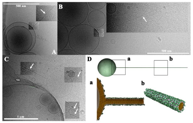
Figure 12.

Cryo-TEM images of block liposomes containing lipid nanorods (white arrows point to nanorods). The block liposomes exist in a narrow composition range (8–10 mol% MVLBG2) of mixtures of the charged lipid MVLBG2 with spherical vesicles of neutral DOPC. A–C: Diblock liposomes (sphere-rod) comprised of lipid nanorods and spherical vesicles. Lipid nanorods are stiff cylindrical micelles with an aspect ratio ≈1000. Their diameter equals the thickness of a lipid bilayer (≈4 nm) and their length can reach up to several microns. D: Schematic of a MVLBG2/DOPC diblock comprised of a lipid nanorod attached to a vesicle. Insets a and b show molecular-scale schematics. Note the high concentration of MVLBG2 in the nanorod. In A–C, image contrast/brightness was altered in selected rectangular areas. Reprinted with permission from [64]. Copyright 2009 American Chemical Society.
