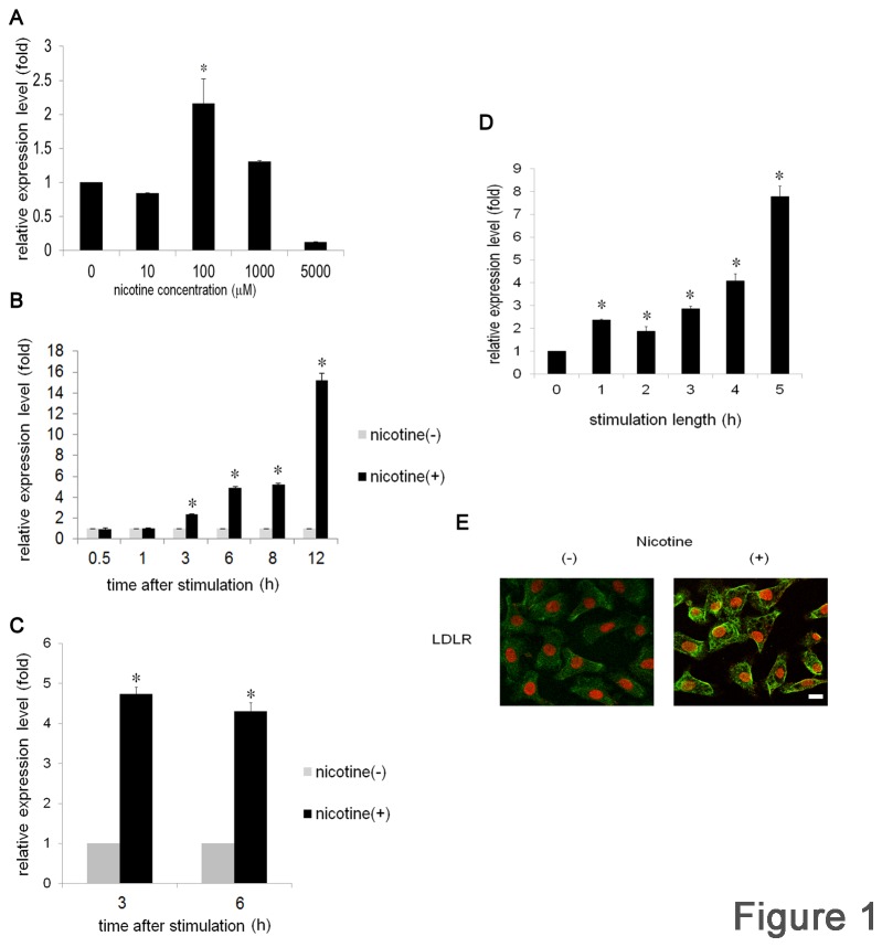
Figure 1. Nicotine induces LDLR expression in OSCC.
Ca9-22 cells were stimulated with various concentrations of nicotine for 3 h (A) or with 100 μM of nicotine for various durations (B). (C) HSC3 cells were stimulated with or without 100 μM of nicotine for 3 and 6 h . (D) Ca9-22 cells were stimulated with 100 μM of nicotine for the indicated time. After stimulation, the cells were washed with PBS, and further cultured with fresh medium for 6 h in total. LDLR mRNA expression levels were examined by real-time PCR. Data from at least 3 separate experiments are shown (mean ± SD). *p < 0.05. (E) Ca9-22 cells were stimulated with or without 100 μM of nicotine for 12 h. Localization of LDLR was detected by immunofluorescence staining with anti-LDLR Ab followed by a FITC-conjugated goat anti-rabbit IgG Ab. Green, LDLR; red, nuclei with monomeric cyanine nucleic acid stain. Scale bar (white line): 10 μm.

